


Dental X-Rays: Illuminating the Path to Optimal Oral Health
Dental X-rays, also known as radiographs, are essential diagnostic tools in dentistry, providing detailed images of teeth, jaws, and surrounding structures invisible during a standard oral exam. With over 90% of U.S. dentists using X-rays annually to detect issues like cavities and bone loss, per the American Dental Association (ADA), these images are critical for early diagnosis and effective treatment. By revealing hidden dental conditions, X-rays enable proactive care, preserving oral health and radiant smiles. This article explores the importance, types, applications, safety, challenges, and future trends of dental X-rays, highlighting their pivotal role in modern dentistry.
The Importance of Dental X-Rays
Dental X-rays provide a comprehensive view of oral structures, uncovering issues not visible to the naked eye. They detect cavities, bone loss, tooth root abnormalities, and impacted teeth, which affect 26% and 50% of U.S. adults for decay and gum disease, respectively, according to the CDC. By identifying problems early, X-rays prevent complications like tooth loss or severe infections, reducing treatment costs by up to 50%. Used in routine check-ups, orthodontic planning, and surgical preparation, X-rays are indispensable for personalized dental care, ensuring both functional and aesthetic outcomes.
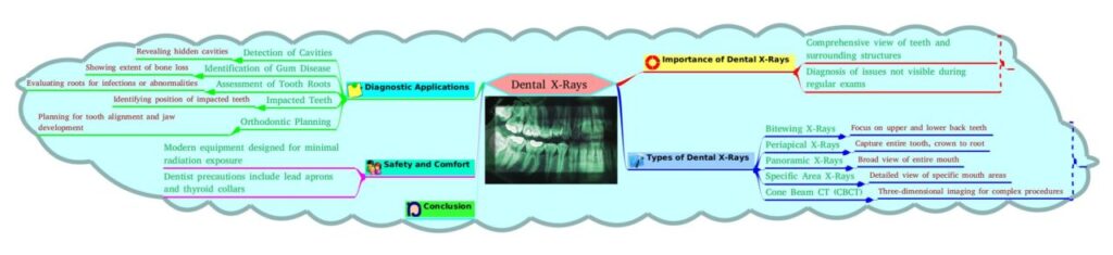
Types of Dental X-Rays
Different X-ray types serve specific diagnostic purposes:
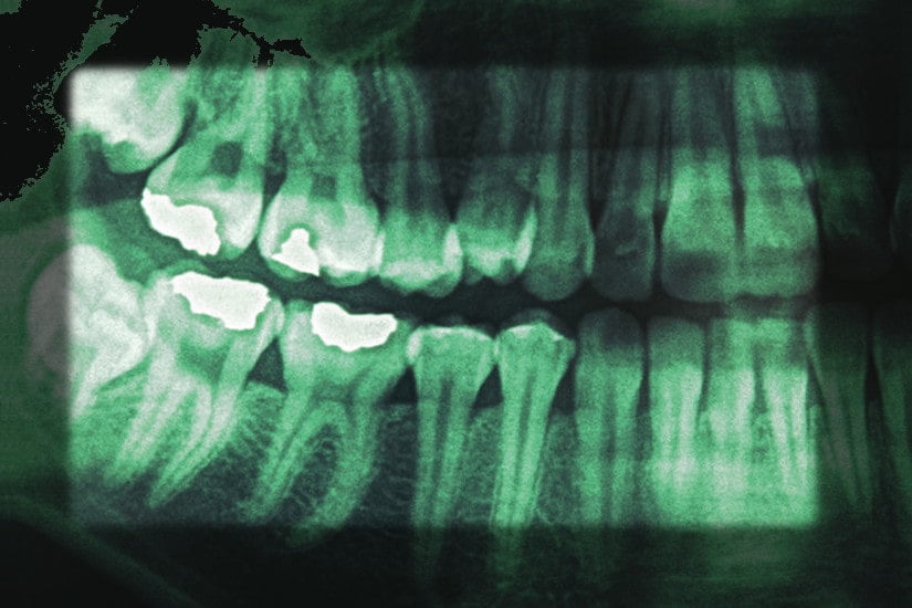
- Bitewing X-Rays: Capture upper and lower back teeth, showing crowns and bone height to detect interdental cavities and early bone loss. Common in routine exams, used every 1–2 years.
- Periapical X-Rays: Image the entire tooth (crown to root) and surrounding bone, identifying root infections, abscesses, or structural damage. Ideal for targeted assessments.
- Panoramic X-Rays: Provide a broad view of the entire mouth, including jaws, teeth, sinuses, and temporomandibular joints (TMJ), used for orthodontics or wisdom teeth evaluation every 3–5 years.
- Occlusal X-Rays: Show specific mouth sections, focusing on tooth development or jaw abnormalities, often used in pediatric dentistry.
- Cone Beam CT (CBCT): Delivers 3D images for complex procedures like implant placement, orthognathic surgery, or TMJ analysis, offering high precision with detailed bone and tissue visualization.
Diagnostic Applications
Dental X-rays support a wide range of diagnoses:
- Cavity Detection: Reveal hidden cavities between teeth or under fillings, missed in 40% of visual exams, per ADA studies.
- Gum Disease Assessment: Show bone loss extent in periodontal disease, affecting 50% of adults, guiding treatment plans.
- Tooth Root Evaluation: Identify infections, fractures, or cysts at the root level, critical for root canal decisions.
- Impacted Teeth: Locate wisdom teeth or other impacted teeth, informing extraction plans in 60% of third molar cases.
- Orthodontic Planning: Assess tooth alignment, jaw development, and crowding, essential for 20% of orthodontic patients.
- Implant and Surgical Planning: CBCT ensures precise implant placement or jaw surgery, improving success rates by 95%.
- Oral Pathology: Detect cysts, tumors, or precancerous lesions, enabling early intervention.
Ensuring Safety and Comfort
Modern dental X-rays prioritize safety:
- Low Radiation: Digital X-rays reduce radiation exposure by 80–90% compared to traditional film, with a single bitewing exposing less than a day’s background radiation.
- Protective Measures: Lead aprons and thyroid collars shield patients, minimizing exposure to vital organs.
- Patient Comfort: Non-invasive and painless, X-rays involve brief positioning of sensors or machines, taking seconds to minutes.
- Special Considerations: Pregnant women avoid routine X-rays, especially in the first trimester, but emergency X-rays use extra precautions (e.g., double shielding).
Challenges and Considerations
Challenges include:
- Cost: X-rays cost $20–$150 (bitewing) to $300–$600 (CBCT), though insurance often covers diagnostic imaging.
- Radiation Concerns: Though minimal, cumulative exposure concerns some patients, requiring clear dentist communication.
- Access: Rural areas may lack advanced imaging like CBCT, affecting 10% of U.S. patients.
- Interpretation: Accurate diagnosis depends on dentist expertise, as misinterpretation can occur in 5% of complex cases.
- Patient Anxiety: Fear of radiation or discomfort, affecting 15% of patients, may require reassurance or sedation for sensitive individuals.
Future Trends
Dental X-ray technology is advancing:
- AI Integration: Artificial intelligence enhances image analysis, improving cavity detection accuracy by 20%.
- Ultra-Low-Dose Imaging: Next-generation digital X-rays further reduce radiation, enhancing safety.
- Portable Devices: Compact X-ray units increase access in underserved areas.
- 3D Imaging Enhancements: CBCT advancements provide higher resolution for precise surgical planning.
- Eco-Friendly Solutions: Digital systems reduce chemical waste from traditional film processing.
Frequently Asked Questions (FAQs)
- How often should I get dental X-rays?
Frequency depends on age, oral health, and risk factors. Adults typically need bitewing X-rays every 1–2 years; high-risk patients may require more frequent imaging. - Are dental X-rays safe?
Yes, modern X-rays use minimal radiation, equivalent to a few hours of natural exposure. Protective gear ensures added safety. - Can pregnant women have dental X-rays?
Routine X-rays are avoided during pregnancy, especially in the first trimester. Emergency X-rays use extra shielding to protect the fetus. - Do dental X-rays hurt?
No, X-rays are painless, involving only brief positioning of sensors or machines. - Why are X-rays necessary if I have no symptoms?
X-rays detect hidden issues like early cavities or bone loss, preventing severe complications in 30–40% of asymptomatic patients.
Conclusion
Dental X-rays are indispensable for diagnosing and managing oral health conditions, from cavities to impacted teeth. By providing detailed insights into hidden structures, they enable early intervention, reducing treatment costs and complications. With low radiation, protective measures, and advancements like AI and 3D imaging, X-rays ensure safe, precise care. Patients should consult their dentist or visit American Dental Association to understand X-ray needs and maintain a healthy smile.